|
Press release on Application of Self Propelling Capsule Endoscope (Fin Type) to Human
June, 2011
|
N. Ohtsuka, President & CEO, MU Ltd., Emeritus Prof., Ryukoku Univ.
Team of Prof. H. Nishihara, Dept. of Mechanical and Systems Eng., Ryukoku Univ.
Team of Prof. K. Higuchi, Internal Medicine(II), Osaka Medical College
|
|
| ABSTRACT |
◇Date: June 21, 2011 (Tuesday) From 10:30 to 11:30
◇Presentors:Naotake OHTSUKA, President, Mu Ltd., Emeritus Prof. of Ryukoku
Univ.
Prof Hironori NISHIHARA and Lecturer Yasunori SHINDO, Ryukoku Univ.
Prof. Kazuhide HIGUCHI and Assoc. Prof. Eiji UMEGAKI, Osaka Medical College
|
| Background |
The capsule endoscope (CE) is an innovative device for painless examination
of the interior of the gastrointestinal tract, especially small intesine,
which was given initial approval by the U.S. Food and Drug Administration
in 2001. More than one million patients worldwide have benefited from the
PillCam SB video capsules (11mm x 26mm) developped by Given Imaging.
Unlike the conventional esophago-gastroduodenoscope (EGD) and colonoscope,
CE
does not allow examiners to observe a
lesion from a desired location and direction in real time. To overcome this disadvantage, we have been endeavoring to develop a microactuator driving system for a capsule endoscope using a magnetic field.
We previously reported successful real-time endoscopic examination of a dog stomach using this driving system. We here report the successful deployment of the SPCE in vivo in stomach
and colon of a human, which suffered no apparent harmful effects on health. |
|
Outline of the system |
Unlike the tube endoscope, a capsule endoscope (CE) occasionally does not
allow examiners to observe a lesion at the desired position and direction
in real time. To overcome this disadvantage, we have developed a self-propelling
capsule endoscope (SPCE), which is propelled in the digestive tract under
the influence of the magnetic field. When a small magnet is placed in an
alternative magnetic field, the magnet vibrates. Then the vibration is
transmitted to a fin and an impelling force is generated (see the animation
in Fig.1).
Using this principal, the improved SPCE has a flexible silicone fin with a small magnet attached at the end of an existing CE and is propelled
by a fluctuating magnetic field remotely.
The moving velocity and direction are controlled freely using a joystick
by adjusting the wave form of the electric current running in the magnetic
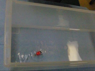
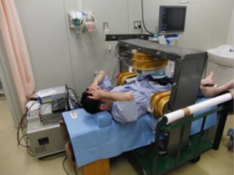
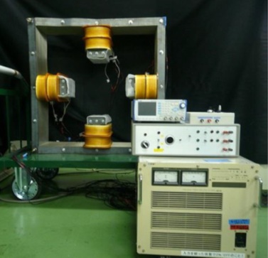
coils. The construction of the SPCE system is shown in Photo1. We gave
a nickname for Mini Mermaid (MM1) to the SPCE in Photo2, the size of which
is 12mm in diameter and 45 mm in total length.
This system was used for examination of the digestive tract in real time.
|
|
|
|
 |
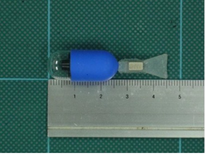 |
| Photo1 Outline of the controlling system for SPCE |
Photo2 Self-propelling capsule endoscope for stomach (MM1) |
|
Summary of in vivo observation of a human:Stomach |
| ●Method of examination |
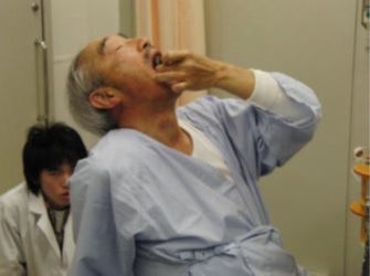
The improved SPCE, the MM1, was swallowed by a human volunteer and propelled
into the stomach of the human for gastric examination in vivo.
Before swallowing the MM1, 500 ml of water was consumed by a volunteer.
The fornix and gastric body were observed at the left decubitus position, the gastric body and gastric angle at the decubitus dorsal position, and the antrum at the right decubitus position.
|
|
|
|
| ●Results of examination in vivo |
(1) The SPCE (MM1) could be swallowed easily and safely without sedation,
and it passed through the esophagus and cardia in a short time (see Photo
4 and movies).
(2) The MM1 could propel by itself under water in the stomach without injuring
the gastric wall, and the moving direction and velocity were controlled
remotely.
(3) Each gastric position could be observed, and the images were obtained
and monitored in real time (see Photo 5 and movies).
|
|
|
|
|
|
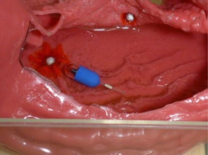
Summary of in vivo observation of a human:Colon |
●Method of examination
A human volunteer stayed at the left decubitus and was injected 500 ml of water passing
anus. The new SPCE (Tall Mermaid, TM1; the total size with 12×65 mm) went
to his left side colon through his anus (see Photo 6 and Photo 4(1)).
The following experiments were conducted: (1) Operation of the SPCE by
remote control in human colon. (2) Obtaining of endoscopic images using
a real time monitoring system.
|
|
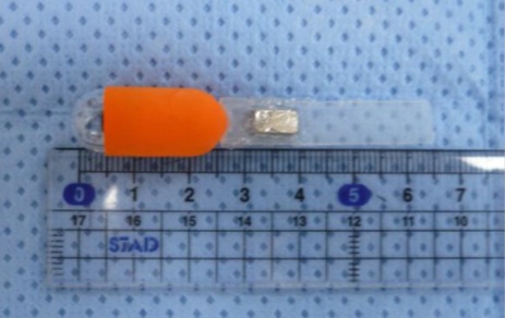 |
Photo6
Self-propelling capsule endoscope for colon(TM1) |
|
| ●Results of examination |
|
(1) The operator was able to drive the SPCE, TM1, in colon by remote control
and it could take clear images .
(2) We could confirm by colon scope that the TM1 moved smoothly in colon.
(3) The TM1 was able to exhaust from his anus easily and safety.
|
|
 |
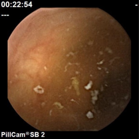 |
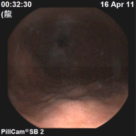
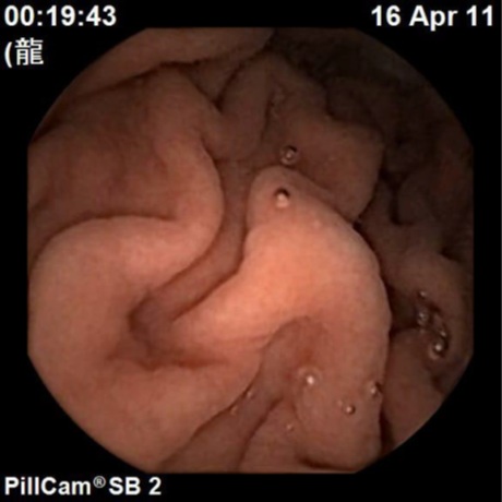
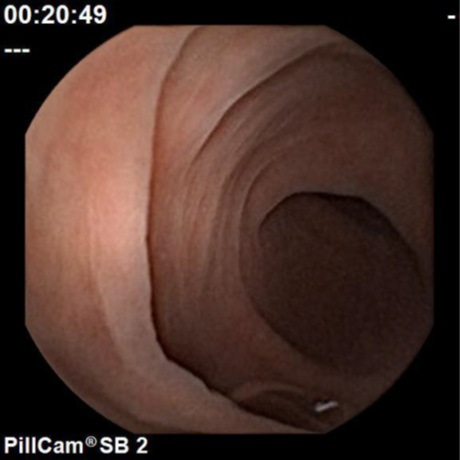
| (1) Sigmoid colon |
(2) Discending colon |
 |
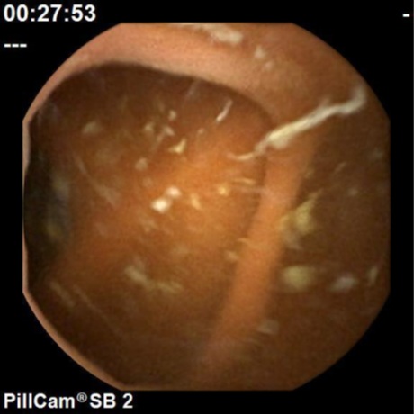 |
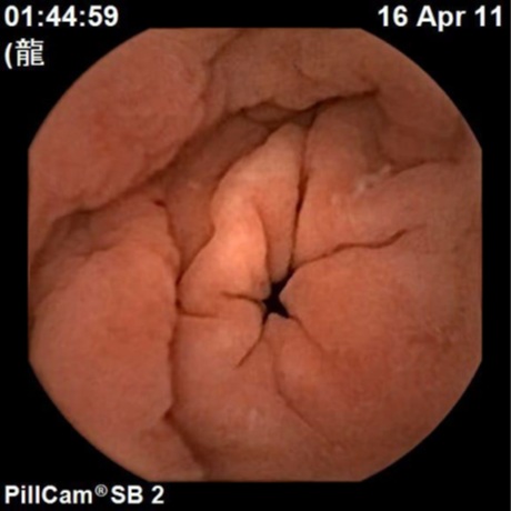
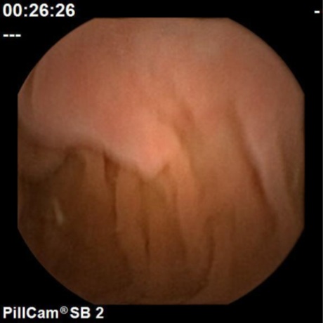
| (3) Spleen curvature |
(4) Transverse colon |
| Photo7 Images of colon shot by TM1 |
|
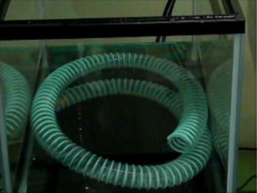
Summary of exhibition:Experiment in models
Exhibition of movement of the MM1 in a phantom stomach and a tube simulating
the small intestine was shown (see Photo 8 and movies). |
|
|
|
Conclusion |
|
The human stomach was examined using an improved SPCE, MM1, safely.
Also we succeeded in observing human colon with SPCE the first time in
the world.
These results suggest that the goal for application of the SPCE to clinical
diagnosis of whole digestive tract would not be far.
|
|